Saturday, December 30, 2006

active transport

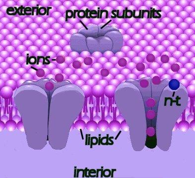
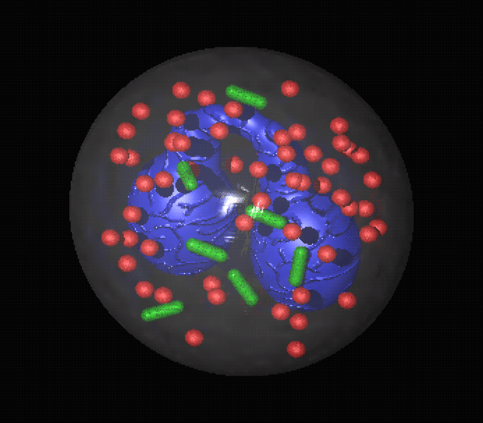
Active transport pumps require expenditure of energy, most often in the form of ATP, to transport hydrophilic macromolecules and ions across membranes against chemical gradients. The number of integral protein transporters in the membrane limits active transport.

Primary, or direct transport involves an energy expending conformational change in the membrane protein to transport a specific molecule or ion across the membrane. Secondary, or indirect transport utilizes energy directly to generate a transmembrane gradient down which ions move and then up or down which the coupled molecule or ion of interest is transported indirectly.

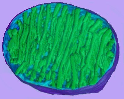
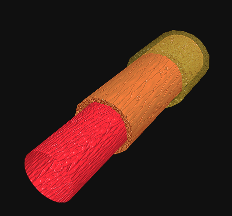
ATPases couple the hydrolysis of ATP (to ADP and Pi) with the transmembrane transport of ions against a concentration gradient. An example is Na+K+ ATPase, which pumps 3 Na+ ions out of the cell for 2 K+ ions it pumps into the cell. Because the pump moves ejects three Na+ for every two K+ moved inward, it generates a net electrical differential necessary for polarization. This electrical potential energy is essential for neuronal activity, and it supplies the energy needed for other types of transport such as symport and antiport. animation - Na K ATPase

Active transport of some substances against a concentration gradient employs the ATPase-derived energy stored in ion gradients, such as proton (H+) or sodium (Na+) gradients, to drive transporter membrane proteins. In a symport, the ATPase transported molecule and the coupled, co-transported ion move in the same direction. Conversely, the ATPase transported molecule and the coupled, co-transported ion move in the opposite direction in an antiport. animation - Na glucose symport

The Virtual Cell Textbook - Cell Biology : Main page of BioChemistry : Main page of Molecules : Main page of Pathways : Main page of Genes : Main page of Cell : Main page of Cell to Cell : Main page of Neuron: Main page of Brain:

apoptosis

Cellular death-by-suicide is part of normal development, and is termed apoptosis or programmed cell death (PCD). Cysteine Aspartate Specific ProteASEs -- caspases -- are active in apoptosis, as is p53, a tumor suppressor gene.
More plus video

Apoptosis and CaspaseGenome Biology Full text DNA-damage signaling and apoptosis: "Cytochrome c binds the apoptosis-activating factor 1 (Apaf1) protein, leading to oligomerization of Apaf1 and caspase 9 into a large 'apoptasome', which then initiates a cascade of caspase activation. Although some non-caspase targets of caspase activation are known, the consequences of proteolysis of these targets are not well understood. Similarly, the events upstream of activation of the caspase cascade in response to DNA damage are not well known; in particular, it is not clear what regulates the decision to undergo apoptosis or to arrest cell proliferation and repair the damage."

apoptosis: Bcl-2 proteins Apoptosis - Bcl-2 proteins: "The bcl-2 proteins are a family of proteins involved in the response to apoptosis. Some of these proteins (such as bcl-2 and bcl-XL) are anti-apoptotic, while others (such as Bad or Bax) are pro-apoptotic. The sensitivity of cells to apoptotic stimuli can depend on the balance of pro- and anti-apoptotic bcl-2 proteins. When there is an excess of pro-apoptotic proteins the cells are more sensitive to apoptosis, when there is an excess of anti-apoptotic proteins the cells will tend to be less sensitive."Cysteine Aspartate Specific ProteASEs -- caspases -- are active in apoptosis, as is p53, a tumor suppressor gene.

Kimball's Apoptosis Page : Apoptosis - Website : Apoptosis Website : Wikipedia on apoptosis : pathology : Cell Suicide in Health and Disease Scientific American Digital : Google apoptosis : animation - zeiosis : Apoptosis, Bcl2, Mitochondria~click on Q for animation : Apoptosis~time-lapse movie : art~apoptosome formation : art~inactivation of DNA repair enzymes : Cell Death Pathways - diagram
Thursday, December 28, 2006

cell membrane

Cell membranes provide adjustable barriers between the cell and the extracellular environment (ESF) or adjacent cells in eukaryotes. The membranes of cellular organelles provide functional compartmentalization from the cytosol. Thus, the cell itself is surrounded by the plasma membrane, and specific functional membranes form intracellular organelles (endoplasmic reticulum, Golgi apparatus) or isolate the contents of cellular organelles (endosomes, mitochondria) from the cytoplasm. art - cell membrane translucent :
art - cell membrane opaque :

Cell membranes are variably constituted of carbohydrates (adhesion and address loci), phospholipid bilayers (hydrophobic barriers), and proteins, which control permeability and cellular signalling. Peripheral membrane proteins are confined to the surfaces of membranes while integral membrane proteins are embedded in the membrane and may pass through the lipid bilayer one or more times.

Specialized membrane proteins function in cell adhesion (junctions) and as energy transducers, enzymes, ion channels, pumps, and receptors for neurotransmitters and hormones. Cell junctions utilize proteins that anchor cells together (desmosomes), that occlude passage of water between cells (tight junctions), and that permit direct communication between cells (gap junctions). diagram . desmosome : tem_desmosome : art - tight junction zonula adherens desmosome gap : diagram . tight junction : diagram . gap junction

ball-stick - globular proteins in phospholipid bilayer : ball-stick - carrier proteins : ball-stick - marker protein : ball-stick - marker proteins : ball-stick - receptor proteins : ball-stick - ion channel proteins: animation - carrier proteins : animation - receptor protein : animation - cholesterol

Main page of Cell : energy transducers : ion channels : pumps : receptors : Main page of Cell to Cell : cellular signalling : GPCRs : GPCR families : Mechanism For Degradation Of G Proteins : neurotransmission : Main page of BioChemistry : enzymes : lipids : neurotransmitters : proteins : phospholipids : receptors : Main page of Molecules : Main page of Pathways: Main page of Genes : Main page of Neuron: synapse :

centriole

Centrioles organize the spindle apparatus on which the chromosomes move during mitosis. Cilia and flagella are organized from peripheral centrioles (basal bodies).

Centrioles consist of 9 sets of triplet microtubules, and centrioles are arranged in pairs perpendicular to each other (tem - 9 triplet pair). animation - spinning centriole pair : tour centriole : zoom in on centriole. Unlike cilia and flagella, which are organized from microtubule organizing centers (basal bodies) at the cell periphery, centrioles have no central doublet of microtubules. Centrioles replicate autonomously, beginning from centers that contain proteins needed for their formation (tubulin, etc.). Procentrioles form first, each erecting a single microtubule from which the triplet can form (diagram - centriole formation). After a single centriole is constructed, daughter centrioles grow out from the tubules at right angles. In a non-dividing cell, they move to the periphery to form the basal body for the cilium (tem - basal bodies). In a dividing cell, the second centriole moves to the daughter cell (in a dividing cell). Where spindles are essential for chromosomal separation during reproduction, cilia are essential for cellular differentiation during embryologic development.

Virtual Cell Textbook - Cell Biology : Cilia, Flagella, and Centrioles : Main page of BioChemistry : Main page of Molecules : Main page of Pathways: Main page of Genes : Main page of Cell : Main page of Cell to Cell : Main page of Neuron: Main page of Brain:

chloroplast

The chloroplast is the site of photosynthesis in eukaryotic cells.

The thylakoid membrane, with its embedded photosystems, is the structural unit of photosynthesis. Both photosynthetic prokaryotes and eukaryotes possess membranes with embedded photosynthetic pigments. Only eukaryotes, which have a nuclear membrane and membrane-bound organelles, possess chloroplasts with an encapsulating membrane. The chloroplast has three compartments, while the mitochondrion has only two. Compartments within a chloroplast are the intermembranous space [3], the stroma [6], and the thylakoid lumen (8) within stromal and granal thylokoids [4,5].

1. outer membrane
2. inner membrane
3. intermembranous space
4. stromal thylakoid
5. granal thylakoid
6. stroma (cytosol)
7. granum (a stack of thylakoids)
8. internal lumen of granal and stromal thylakoids
(click to enlarge image)

The typical higher plant chloroplast is lenticular and approximately 5 microns at its largest dimension. Plant cells contain from 1 to 100 chloroplasts, depending on the type of cell. The mature chloroplast is typically bounded by outer (1) and inner (2) membranes that possess significantly different chemical constituents. In addition to enzymes that function in photosynthesis, chloroplasts also contain a circular DNA molecule and the protein-synthetic machinery characteristic of prokaryotes.

Each chloroplast contains about 40 to 80 grana (7), and each grana comprises about 5 to 30 thylakoids. The thylakoids are membranous disks about .25 to .8 microns in diameter, which contain protein complexes, pigments, and other accessory components. The phospholipid bilayer of the thylakoid is folded repeatedly into stacks of grana. (details) These stacks are connect by channels to form a single functional compartment.

The thylakoid is the site of oxygenic photosynthesis in eukaryotic plants and algae, and in prokaryotic Cyanobacteria. Cyanobacteria possess thylakoid membranes, but as prokaryotes they do not contain chloroplasts. Chlorophyll, accessory pigments, and other integral membrane proteins transduce light energy to provide excited electrons (excitons) to electron transport chains, powering the formation of NADPH and ATP during photophosphorylation.As intracellular plant organelles, chloroplasts are classified as plastids.

Chloroplasts originate within the eukaryotic photosynthetic cell either by division of pre-existing plastids or from protoplastids (proplastid). These proplastids are organelles with little internal structure, enclosed within two dissimilar membranes. It is assumed that thylakoid membranes form during the chloroplast maturation process and are derived from the inner membrane of the proplastid and chloroplast.

The current consensus is that chloroplasts originated from Cyanobacteria that have become endosymbionts. This is an origin analogous to the endosymbiotic origin of mitochondria, which are believed derived from "purple bacteria", alpha-proteobacteria most closely related to Rickettsiales.

cilia

The primary cilium is a slender protuberance on the surface of nearly every cell in the body. Cilia are functioning organelles know to be essential to normal development and health. Some cilia are rigid spikes that gather sensory information, while other cilia are flexible and whip-like, registering or directing flow in the surrounding fluid. Cilia and flagella both have an internal structure built upon microtubules, but the flagellum is longer and is more often a single organelle. Inside both cilia and flagella is a microtubule-based cytoskeleton termed the axoneme, which provides scaffolding for various protein complexes. 3D diagram - axoneme : 3D animation – inside flagellum : image - detail of cilia : tem - structure cilium : diagram - mechanism of ciliary motility : Geometric Clutch Model : animation - cilia & flagella

The motor protein dynein powers the sliding of the microtubules against one another — first on one side, then on the other. This dynein-powered sliding produces a whip-like motion employed to move fluid past or over the cell. During the course of evolution, cilia have been adapted to function as mechanoreceptors, chemoreceptors, and the outer segment of the rods in the vertebrate retina. Although called stereocilia, the hair-cell protrusions in the inner ear are actually modified villi. sem image - inner ear stereocilia

The microtubular axoneme also provides binding sites for molecular motor proteins, such as kinesin II, which assist in the transport of proteins up and down the microtubules. 3D diagram - axoneme The microtubule organizing center, also called a basal body, lies at the base of the cilium. tem - basal bodies. The basal body is created as the centriole (a microtubular structure essential to cell division) migrates to the surface. The transition zone between axoneme and basal body serves as a docking station for intraflagellar transport and motor proteins. During intraflagellar transport (IFT) materials needed to build the cilia are carried to the ciliary tip and spent materials are carried down to the ciliary body. The IFT particle, which is made up of at least 17 polypeptide subunits, may also carry signals collected by various receptors embedded in the ciliary membrane.

More: Cilia, Flagella, and Centrioles : Cilia and flagella :
HHMI Bulletin September 2005: The Importance of Being Cilia
Google cilia

Virtual Cell Textbook - Cell Biology : Main page of BioChemistry : Main page of Molecules : Main page of Pathways : Main page of Genes : Main page of Cell : Main page of Cell to Cell : Main page of Neuron: Main page of Brain:

cytoplasm

Cytoplasm fills cells from the nuclear membrane to the plasma membrane, and is composed of fluid cytosol (or viscid cytogel) and the cellular organelles. Eighty percent of cytoplasm is cytosol, or hyaloplasm, the aqueous component that contains ions, carbohydrates, salts, enzymes, proteins, and RNA. Cellular organelles pack the cytoplasm – centrioles, microtubules of the cytoskeleton, endoplasmic reticulum, endosomes, exosomes, the Golgi apparatus, mitochondria, and peroxisomes. clickable art - cell : clickable diagram - animal cell

Virtual Cell Textbook - Cell Biology : Main page of BioChemistry : Main page of Molecules : Main page of Pathways :Main page of Genes : Main page of Cell : Main page of Cell to Cell : Main page of Neuron: Main page of Brain

cytoskeleton

The cytoskeleton is a dynamic three-dimensional filamentous structure within the cytoplasm of eukaryotic cells. It is constructed of microfilaments, intermediate filaments, and microtubules that provide for structural support and movement. image - cytoskeleton : image_cytoskeleton : diagram - mechanism of ciliary motility .

Microfilaments are 3-6 nm in diameter, and are composed mostly of the contractile protein actin – the most abundant cellular protein. Microfilaments are responsible for the cellular movements of gliding, contraction, and cytokinesis. The association of micofilaments of actin with the protein myosin is central to muscle contraction. image_filamentous actin microtubules nuclei : image_filamentous actin & microtubules : image_microtubules nuclei endothelial tc : image_filamentous actin microtubules nuclei fibroblast mouse : image_tubulin microtubules.

Intermediate filaments are about 10 nm diameter and provide tensile strength for the cell connect adjacent cells through desmosomes (macula adherens). diagram . desmosome : tem_desmosome : diagram . tight junction : diagram . gap junction :

Virtual Cell Textbook - Cell Biology : Main page of BioChemistry : Main page of Molecules : Main page of Pathways :Main page of Genes : Main page of Cell : Main page of Cell to Cell : Main page of Neuron: Main page of Brain
Wednesday, December 27, 2006

differentiation

Tuesday, December 26, 2006

energy transducers

An energy transducer transforms one mode of energy to another.

For example, for most sensory modalities sensory cells act as energy transducers, transforming the energy of the environmental stimulus into a change in the electrical potential difference across a biological membrane. Bacteriorhodopsin (bR) is an integral protein in the plasma membrane of a bacterium. Upon light absorption, bR transports protons across the membrane, converting the photon energy into the energy of a proton electrochemical gradient. bR is a single small protein and is the simplest known active ion pump and biological light energy transducer. Consequently, bR is a prototype system for studying the basic steps and rules of biological energy transduction. Energy transduction is thus the fundamental physical basis of the sensory response of most biological systems to their environments. Within each of the evolved sensory modalities there exists an enormous variety of structure and function produced by evolutionary pressures. Rhodopsins are found in one of the GPCR families.

Oxidative phosphorylation within the mitochondrion is another form of energy transduction, as is the conversion of chemical energy to muscle contraction and relaxation.

Virtual Cell Textbook - Cell Biology : Main page of BioChemistry : Main page of Molecules : Main page of Pathways :Main page of Genes : Main page of Cell : Main page of Cell to Cell : Main page of Neuron: Main page of Brain

endoplasmic reticulum

image gjh.md
1. smooth endoplasmic reticulum (smooth ER)
2. cisternal space -- lumen -- of rough endoplasmic reticulum (rough ER) with proteins produced on exterior
3. cisternae of rough ER
4. chains of ribosomes along mRNA on membrane of rough ER, the proteins move into the lumen of the cisterna following assembly
5. rosette-shaped nuclear pore (size exaggerated)
6. nuclear membrane continuous with membrane of ER
7. strands of heterochromatin adherent to nucleolus
8. nucleolus, site of production of ribosome components
9. nuclear material in nucleus (cut-away)

endosomes

UCSB Researchers Discover That The Cell's Endosomes Use A Surprising Transportation System: "Endosomes travel to the cell's nucleus using back-and-forth symmetrical movement, rather than taking a more direct route. This forward and reverse motion leads to even distribution of the endosomes on microtubules.

An aster-like layout of the microtubules helps the endosomes accumulate at the nucleus. The researchers think this non-direct approach to the nucleus has evolved to allow hundreds of endosomes to bring nutrients and molecular information to the cell's center for processing. Even if the cell moves or if there's increased traffic flow, there's never a traffic jam on the microtubules.

While it has long been known that endosomes travel in a bidirectional way, it has not previously been established that the transport system is symmetrical."

Virtual Cell Textbook - Cell Biology : Main page of BioChemistry : Main page of Molecules : Main page of Pathways :Main page of Genes : Main page of Cell : Main page of Cell to Cell : Main page of Neuron: Main page of Brain

eukaryotic

Virtual Cell Textbook - Cell Biology : Main page of BioChemistry : Main page of Molecules : Main page of Pathways :Main page of Genes : Main page of Cell : Main page of Cell to Cell : Main page of Neuron: Main page of Brain

exosome

The exosome complex consists almost exclusively of exoribonucleolytic proteins -- 10 different proteins important for 3'– > 5' degradation of ARE-containing mRNAs in mammalian cells. Although exosomes accumulate in the nucleolus, they also localize in the cytoplasm and in neoplasm. The exosome membrane is organized as a lipid bilayer with a random distribution of phosphatidylethanolamines. Exosome membranes display a similar content of the major phospholipids and cholesterol, but an enrichment in sphingomyelin when compared to the parent cell membrane.

Review Exosome & Models of exosome complex.

Edinburgh Research Archive : Item 1842/734: "The exosome complex of 3' -> 5' exoribonucleases functions in both the precise processing of 3' extended precursor molecules to mature stable RNAs and the complete degradation of other RNAs. Both processing and degradative activities of the exosome depend on additional cofactors, notably the putative RNA helicases Mtr4p and Ski2p. "

Virtual Cell Textbook - Cell Biology : Main page of BioChemistry : Main page of Molecules : Main page of Pathways :Main page of Genes : Main page of Cell : Main page of Cell to Cell : Main page of Neuron: Main page of Brain
Sunday, December 24, 2006

Golgi apparatus

image gjh.md

The Golgi apparatus (purple) is bound by a single membrane, and is similar to the endoplasmic reticulum (blue). The apparatus comprises a stack of large membrane-bound vesicular plates (4) important in packaging macromolecules for transport within the cell. The cis side of the Golgi complex is closer to the ER, and the trans side is farther from the ER. Golgi bodies are vesicles that have bud off (3) from the plates. animation - golgi apparatus and budding golgi body : animation - golgi at work : animation - lysosomes "suicide sacks" : tour lysosome

Thus, the stack of large vesicles is surrounded by numerous small vesicles (2) containing the macromolecules . The hormonal, neurotransmitter, or enzymatic content of lysosomes, peroxisomes and secretory vesicles are contained in these membrane-bound vesicles at the periphery of the Golgi apparatus. Delivery of macromolecules is effected by symmetric back-forth endosomal transport along the microtubules of the cytoskeleton.

Virtual Cell Textbook - Cell Biology : Back to: Main page of BioChemistry : Main page of Cell : Main page of Genes :
Main page of Neuron: synapse : dendrite : Main page of Brain:

Friday, December 22, 2006

ion channels

image gjh.md

Ion channels undergo a conformational change -- an alteration of 3D shape -- when coupled with a neurotransmitter (n-t). This conformational change of the protein subunits enlarges the channel pore, permitting entry of ions to the interior of the cell.

The cell membrane comprises a phospholipid bilayer membrane with hydrophobic ends oriented toward the abutting phospholipid molecule. image - cell membrane cross-section : animation - phospholipid : image - bilayer of phospholipids in aqueous solution. The hydrophilic ends of the two layers are oriented toward the exterior or toward the cell's cytoplasm. Various proteins, including the subunits of ion channels, are embedded in the membrane. ball-stick - globular proteins in phospholipid bilayer : ball-stick - ion channel proteins : animation - facilitated diffusion.

Los Alamos National Laboratory (LANL): Scientists propose new method for studying ion channel kinetics: "Faulty ion channels in humans have been shown to cause severe diseases like cystic fibrosis and diabetes and more subtle, but still dangerous physiological effects, like over-responses to general anesthetics. "Ions such as calcium, sodium and potassium play a fundamental role in nearly all biological processes. Calcium, for example, is important in fertilization, cell death, cell division, human hearing, memory, vision, and the immune system. It also plays a factor in cancer, Alzheimer's, alcohol caused neuronal damage, migraine headaches, cardiomyopathy (heart failure), hypertension and a host of other normal and abnormal physiological functions. Other ions and ion channels are important for processes such as muscle contraction and nerve conduction.""

Virtual Cell Textbook - Cell Biology : Main page of BioChemistry : Main page of Molecules : Main page of Pathways :Main page of Genes : Main page of Cell : Main page of Cell to Cell : Main page of Neuron: Main page of Brain

Tuesday, December 19, 2006

lysosome

Monday, December 18, 2006

microtubules

Microtubules are cylindrical tubes 20-25 nm in diameter, which are composed of linear polymers (protofilaments) of the globular protein tubulin. The tubulin molecules form heterodimers of alpha and beta tubulin and linear rows of tubulin dimers form the protofilaments. Microtubules act as scaffolding to maintain cell shape, and extend throughout the cytoplasm of eukaryotic cells. image_filamentous actin microtubules nuclei : image_filamentous actin & microtubules : image_microtubules nuclei endothelial tc : : image_filamentous actin microtubules nuclei fibroblast mouse : image_tubulin microtubules

In addition to this cytoskeletal role, microtubules act as cellular conveyer belts, employing special attachment proteins to move chromosomes, granules, endosomal vesicles, and organelles such as mitochondria through the cytoplasm.

Microtubules may work alone, or be joined with other proteins to form more complex structures such as cilia and flagella and centrioles. Within cilia and flagella, microtubules are assembled in a 9 + 2 arrangement. animation - inside flagellum. The tubulin is coupled to dynein arms to enable locomotion (spermocytes, protozoa) or movement of liquid over the cell (important in embryologic differentiation). image_spermatozoa (mouse) : image_sperm (Dv) : diagram - mechanism of ciliary motility : animation - cilia & flagella

Arranged in orthogonal paired tubes of 9 fibers, microtubules form centrioles during cell division, or basal bodies at the root of cilia and flagella. animation - spinning centriole pair : tour centriole : zoom in on centriole. During mitosis microtubules form spindle fibres along which chromosomes assemble then separate. animation - mitosis : animation ~ mitosis : link to animation - mitosis : kyrk animation _ mitosis : image_mitosis microtubules kinetochores DNA : image_mitosis : image_aberrant division mammalian cell : image_anaphase : image_golgi apparatus DNA microtubules dividing cells : image_mitotic spindle : image - spindle : animation - mitosis animation - meiosis : kyrk animation _ meisosis : Google cytoskeleton : Google microtubule

Virtual Cell Textbook - Cell Biology : Main page of BioChemistry : Main page of Molecules : Main page of Pathways :Main page of Genes : Main page of Cell : Main page of Cell to Cell : Main page of Neuron: Main page of Brain

mitochondrion

The mitochondrion (pl. mitochondria) is the 'power house of the eukaryotic cell, performing oxidative phosporylation. Mitochondria have two internal, membrane-bound spaces, unlike chloroplasts, which have three internal spaces.

The outer mitochodrial membrane is similar in constitution to the eukaryotic cell’s plasma membrane, while the inner membrane is similar in chemical composition to bacterial membranes. This difference is one of several lines of evidence for the serial endosymbiotic origin of mitochondria as phagocytozed purple bacteria.

Above left – click to enlarge : simplified diagram of a mitochondrion showing:
1. space between inner and outer membranes
2. matrix
3. christae
4. junction between membranes
5. inner membrane
6. outer membrane

The image at right (click to enlarge) is based on reconstruction of serial tem slices through a mitochondrion. The outer membrane (violet) surrounds the organelle. The inner membrane (pale blue) is contiguous, at membrane junctions (pale blue connecting to green at lower center), with the inner membrane that forms the walls of cristae (green).

The matrix – a soup factory – lies between the cristae, and contains mitochondrial DNA and the components of intermediary metabolism. image - mitochondrion cut : tour mitochondrion :

The outer and inner membranes are composed of phospolipid bilayers studded with proteins, much like the cell membrane. However, the composition of the inner and outer membranes is very different.

The inner mitochondrial membrane contains more than 100 different polypeptides. The protein to phospholipid ratio is very high – more than 3:1 by weight, having about 1 protein for 15 phospholipids. The inner membrane is also rich in an unusual phospholipid, cardiolipin, which is usually characteristic of bacterial plasma membranes. This composition, along with other evidence, has led to the assumption that the inner membrane is derived from endosymbiotic prokaryotes. The endosymbiotic theory of eukaryotic evolution is now widely accepted.

In contrast, the outer membrane, which encloses the entire mitochondrion, is similar in composition to the cell membrane and comprises about 50% phospolipids by weight and contains a variety of enzymes. The enzymes carry out activities such as the oxidation of epinephrine (adrenaline), the degradation of tryptophan, and the elongation of fatty acids.

The plant chloroplast is the site of photosynthesis : animation - chloroplast : tour the chloroplast : Virtual Cell Textbook - Cell Biology
Sunday, December 17, 2006

nuclear membrane

nuclear pore

animation - spinning nucleus : art - nuclear membrane and nuclear pores : Virtual Cell Textbook - Cell Biology : Main page of BioChemistry : Main page of Molecules : Main page of Pathways : Main page of Genes : Main page of Cell : Main page of Cell to Cell : Main page of Neuron: Main page of Brain:

nucleolus

animation - nucleolus heterochromatin : animation - spinning nucleus :
art - nuclear membrane and nuclear pores : Virtual Cell Textbook - Cell Biology : Main page of BioChemistry : Main page of Molecules : Main page of Pathways : Main page of Genes : Main page of Cell : Main page of Cell to Cell : Main page of Neuron: Main page of Brain:

nucleus

Friday, December 15, 2006

peroxisome

The peroxisome is a cellular organelle bounded by a single-membrane that is present in most eukaryotic cells. Beta oxidation of long and very long chain fatty acids occurs in the peroxisome. The organelle is also involved in bile acid synthesis, cholesterol synthesis, plasmalogen synthesis, amino acid metabolism, and purine metabolism. The oxidative enzymes in peroxisomes protect the cell from metabolic production of hydrogen peroxide by breaking H2O2 down into water and oxygen.

Biochemistry of the Peroxisome @ Peroxisome.org : Beta oxidation : Auxiliary b-oxidation : a-oxidation : Glycerolipid synthesis : Cholesterol biosynthesis : Amino acid metabolism : Polyamine degradation : Purine metabolism : Bile acid synthesis : Biochemistry of the peroxisome membrane : Peroxisomal matrix enzymes : Peroxisomal single enzyme disorders

Defects in these peroxisomal processes manifest in numerous genetic disorders, which can be divided into two categories: disorders resulting from a defect in a single peroxisomal enzyme, and disorders which result from a deficiency in the biogenesis of the peroxisome, affecting all of the metabolic pathways of the peroxisome. Disorders related to the peroxisome can result from mutations in any of at least 13 genes, known as PEX genes. The products of these genes are known as peroxins, and all function in some aspect of peroxisome biogenesis.

Google peroxisome : The Peroxisome for the Scientist - Physician - Layperson : Main page of BioChemistry : Main page of Molecules : Main page of Pathways : Main page of Genes : Main page of Cell : Main page of Cell to Cell : Main page of Neuron: Main page of Brain:

phagocytosis

Phagocytosis involves the sporadic engulfment of large extracellular particles by wrapping cytoskeletally supported pseudopodia around the particle and internalizing it into vacuoles called phagosomes. The phagosome then fuses with a lysosome, and the phagocytosed particle is digested in an oxidative burst by lysosomal enzymes. The hydrolyzed products are absorbed into the cytoplasm through the vacuolar wall, and the waste products are excreted from the cell. image_Paramecium feeding on hematococcus : animation - phagocytosis : animated diagram - phagocytosis of bacterium : time-lapse movie - phagocytosis : animation - exocytosis

Back to: Main page of BioChemistry : Main page of Molecule : Main page of Pathways: Main page of Genes : Main page of Cell : transport : Main page of Cell to Cell : Main page of Neuron: Main page of Brain:

plant cells

The focus of this site is the animal cell, but for the sake of completeness : image - vacuole plant : image - cell wall plant : animation - spinning plant plastid : clickable image - cell : Virtual Cell Textbook - Cell Biology

Virtual Cell Textbook - Cell Biology : Main page of BioChemistry : Main page of Molecules : Main page of Pathways : Main page of Genes : Main page of Cell : Main page of Cell to Cell : Main page of Neuron: Main page of Brain:

pinocytosis

Pinocytosis is a continuous process in most cells. Pinocytosis is called "cellular drinking" and involves encysting small quantities of extracellular fluid (ESF). View animation - pinocytosis

Back to: Main page of BioChemistry : Main page of Molecule : Main page of Pathways: Main page of Genes : Main page of Cell : transport : Main page of Cell to Cell : Main page of Neuron: Main page of Brain:

Google pinocytosis

protein degradation

The FHA-domain: a common motif found in nuclear kinases and: "The majority of intracellular protein degradation takes place at the proteasome, a multi-subunit protease present in the cytoplasm and the nucleus of eukaryotic cells. The 20S proteasome forms a barrel-shaped structure, with the active sites of the multiple catalytic subunits facing the central cavity. This core proteasome is typically associated with a 19S cap complex, resulting in a larger assembly termed the 26S proteasome. The 19S cap complex contains several ATPase subunits, which probably act in unfolding the target proteins. In addition, the 19S complex contains at least eleven non-ATPase subunits, which are thought to function in regulation and target recognition. One of these subunits (S5a) binds to ubiquitin, which serves as a universal signal for targeting proteins to the proteasome. Two of the regulatory subunits have recently been shown to share a repeat motif with the cyclosome/APC-complex, which acts in the cell cycle dependent ubiquitination of regulatory proteins. In order to learn more about the potential function of the regulatory proteasome subunits, we set out to identify distantly related proteins by applying the generalised profile method, a sensitive motif-based technique for sequence database searches. We were able to identify two different homology domains, which both occur in multiple components of the regulatory proteasome as well as in several other characterised protein families"

prokaryotic

Prokaryotes are microbial cells lacking a nuclear membrane. Eukaryotes are cells whose nuclear material is enclosed by a nuclear membrane.

The Eubacteria of Kingdom Monera, commonly called bacteria:
1. Prokaryotic organisms without a nuclear membrane
2. Eubacterial rRNA and no introns in genome
3. Membrane lipids are primarily diacyl glycerol ethers
4. Cell walls contain thick (gram+) or thin layers of peptidoglycan (gram-)

The Archaea, or Archaeobacteria
1. Prokaryotic organisms without a nuclear membrane
2. Archaeobacterial rRNA -- introns in genome
3. Membrane lipids are unusual -- primarily isoprenoid glycerol diether or diglycerol tetraether derivatives
4. The cell wall does not contain peptidoglycan (gram-)

There are three groups of Archaea, which also called extremophiles: Methanogens are poisoned by O2, Thermophiles live in extreme temperatures, and Halophiles live in highly saline environments

Prokaryote structure : Prokaryote taxonomy & phylogeny : Eubacteria and Archaea : Eubacteria : The Archaea : Virtual Cell Textbook - Cell Biology : Main page of BioChemistry : Main page of Molecules : Main page of Pathways : Main page of Genes : Main page of Cell : Main page of Cell to Cell : Main page of Neuron: Main page of Brain:

pumps

Wednesday, December 13, 2006

receptor

image - receptor proteins : animation - receptor protein

Virtual Cell Textbook - Cell Biology : Main page of BioChemistry : Main page of Molecules : Main page of Pathways : Main page of Genes : Main page of Cell : Main page of Cell to Cell : Main page of Neuron: Main page of Brain:

receptor-mediated endocytosis

Receptor-mediated endocytosis creates receptosomes. Cells invaginate proteins and other types of ligands that have attached to specific receptors on the plasma membrane. First, the protein or ligand binds to a specific receptor, forming a coated pit ("coated pit endocytosis"). The coated pit is a specialized membranous region coated with clathrin, which provides stability and aids the transport process. Next, the coated pit next forms a coated vesicle and, shedding its clathrin coat, joins with other coated pits to form a receptosome.

Back to: Main page of BioChemistry : Main page of Molecule : Main page of Pathways: Main page of Genes : Main page of Cell : transport : Main page of Cell to Cell : Main page of Neuron: Main page of Brain:

Google receptor-mediated endocytosis

ribosome

Ribosomes are large intracellular aggregates attached to the rough endoplasmic reticulum. They comprise several RNAs and scores of proteins, and function as ribozymes.

Ribosome biogenesis in eukaryotes mainly occurs in the nucleolus, a specialized nuclear compartment. The synthesis of rRNAs is not achieved by simple transcription of the individual rRNA species, rather it requires a complex series of post-transcriptional processing steps.

Largest Computational Biology Simulation Mimics Life's Most Essential Nanomachine: "The ribosome is so fundamental to life that many portions of this molecular machine are identical in every organism ever genetically sequenced. In developing the project, the team identified a corridor inside the ribosome that the transfer RNA must pass through for the decoding to occur, and it appears to be constructed almost entirely of universal bases, implying that it is evolutionarily ancient." The original news release can be found here. Voxel simulation image : Image 2 : Image 3 : Image 4 : Image 5 : High res movie : Low res movie : See an image of the Q machine at http:/​/​www.lanl.gov/​asci/​.

Back t0: Main page of BioChemistry : amino acid : catalyst : enzyme : peptide : protein : ribozyme : Main page of Cell : endoplasmic reticulum : Main page of Genes : RNA : microRNA : mRNA : rRNA : tRNA : RNA world : archival DNA : Main page of Brain: Main page of Neuron : neuron
Tuesday, December 12, 2006

spindle

CELL BIOLOGY: ON THE ORCHESTRATION OF THE MITOTIC SPINDLE: "New work provides the clearest evidence yet that spindle assembly is coordinated by the generation, at chromosomes, of an intracellular gradient of the active guanosine triphosphate (GTP)-bound form of Ran, a small GTPase of the Ras super-family present in all eukaryotic cells."
Monday, December 11, 2006

transport

Movement across cell membranes is characterized as passive or active, and is dependent upon the nature of the phospholipid bilayer, ion channels, and protein pumps, which permit simple diffusion, facilitated diffusion, and active transport respectively.

The cell membrane is more permeable to non-polar, hydrophobic molecules than to polar, hydrophilic molecules by virtue of the hydrophobic interior of the amphipathic lipids of the bilayer. As a result, some non-polar molecules such as H2O and CO2 are able to diffuse directly across the cell membrane down a concentration gradient. This osmotic, chemical gradient limits both the rate of diffusion and the maximum concentration of the diffusing molecule in the cytosol, or the extracellular fluid (ESF) in the case of waste products.

Small polar molecules, typically simple ions like H+, K+, Na+, or Cl- are sufficiently small for facilitated diffusion through transmembrane protein pores called ion channels. The rate of facilitated diffusion is determined both by the number of ion channels and the concentration gradient. image - ion channel proteins : animation - facilitated diffusion

Active transport utilizes energy to move molecules or ions against concentration gradients. image - carrier proteins : animation - carrier proteins : animation - Na glucose symport : animation - Na K ATPase

In addition, though not strictly a form of transmembrane transport, molecules can be moved through membranes. Endocytosis involves movement of macromolecules from the exterior into the lumen of endomembranous organelles. This is achieved by phagocytosis, pinocytosis, or receptor-mediated endocytosis. animation - phagocytosis. Exocytosis is the reverse of endocytosis. Vesicles containing the substances for release are transported to the plasma membrane and fuse with it, effecting extrusion of vesicular contents. animation - exocytosis The soluble NSF receptor (SNARE proteins) are the best characterized among many factors involved in exocytosis.

Google endocytosis / Google exocytosis / Google phagocytosis / Google pinocytosis / Google receptor-mediated endocytosis

Virtual Cell Textbook - Cell Biology : Main page of BioChemistry : Main page of Molecules : Main page of Pathways : Main page of Genes : Main page of Cell : Main page of Cell to Cell : Main page of Neuron: Main page of Brain:
Saturday, December 09, 2006

vacuole

animation - contractile vacuoles of protozoa : Virtual Cell Textbook - Cell Biology : Main page of BioChemistry : Main page of Molecules : Main page of Pathways : Main page of Genes : Main page of Cell : Main page of Cell to Cell : Main page of Neuron: Main page of Brain:

vacuole

Saturday, December 02, 2006

vesicle

© The content of this blog is intended for personal or educational use. All rights reserved for commercial use of contents. Unless otherwise specified, all images created by site author.